Researchers have discovered a groundbreaking use of terahertz (THz) imaging to visualise cochlear structures in mice, offering non-invasive, high-resolution diagnostics. By creating 3D reconstructions, this technology opens new possibilities for diagnosing hearing loss and other conditions. THz imaging could lead to miniaturised devices, such as THz endoscopes and otoscopes, revolutionising diagnostics for hearing loss, cancer and more. With the potential to enhance diagnostic speed, accuracy and patient outcomes, THz imaging could transform medical practice.
Advancements in healthcare and technology have significantly increased the average human lifespan. However, with longer life comes a higher prevalence of age-related disorders that affect overall wellbeing. One such condition is hearing loss in older adults, which can severely impact communication, social interactions and daily functioning.
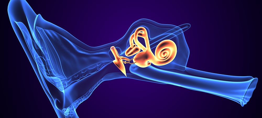
Hearing relies on the cochlea, a spiral-shaped organ in the inner ear that converts sound waves into neural signals. Any structural or functional impairment of the cochlea can lead to hearing loss, making its precise visualisation essential for understanding and diagnosing auditory disorders. Conventional imaging techniques often struggle to capture the intricate details of this delicate structure, necessitating the development of more advanced imaging approaches.
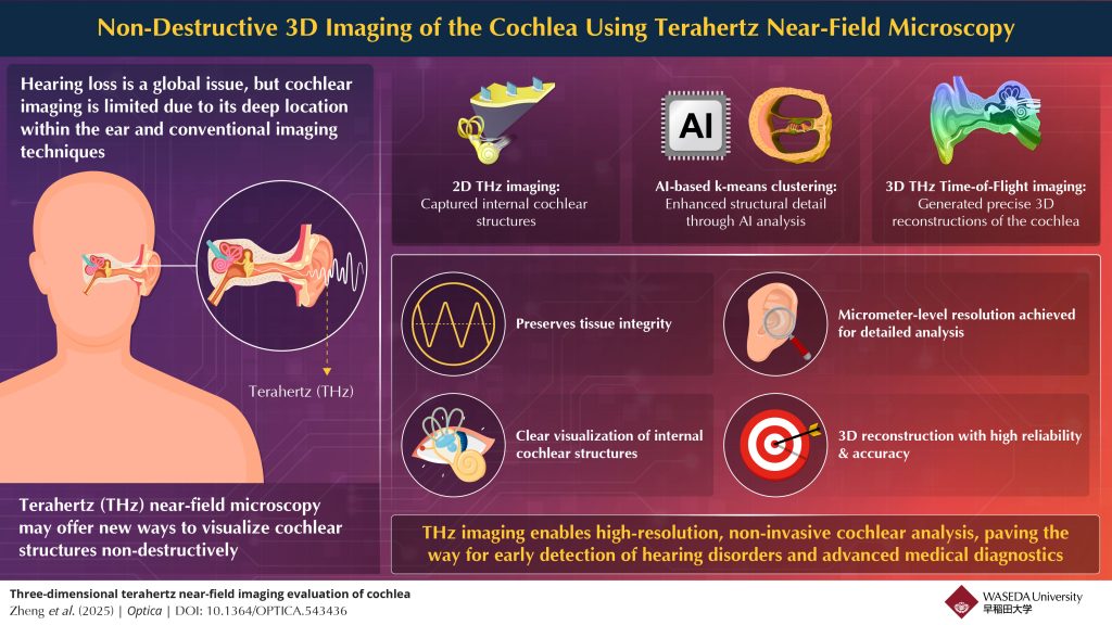
To investigate the potential of terahertz (THz) imaging for visualising cochlear structures, researchers led by Associate Professor Kazunori Serita from Waseda University, along with Professors Takeshi Fujita and Akinobu Kakigi from Kobe University and Professor Masayoshi Tonouchi and Luwei Zheng from Osaka University, used a micrometre-sized THz point source to visualise the internal structure of the mouse cochlea. The study, published in Optica on 27 March 2025, explores THz imaging as a non-invasive, high-resolution technique for biological tissue analysis. “By leveraging THz waves, we can achieve deeper tissue penetration while preserving structural clarity,” explains Serita.
To achieve high-resolution THz imaging, a micrometre-sized THz point source was generated using a femtosecond laser at a wavelength of 1.5 μm, which irradiated a GaAs substrate. The cochlea was placed directly on the substrate to facilitate near-field imaging. The system captured 2D THz time-domain images over a broad timescale, allowing structural visualisation at varying depths. By applying the time-of-flight principle, the timescale of each THz image was converted into a depth scale. Furthermore, k-means clustering, an unsupervised machine-learning technique, was used to extract structural features and enable 3D reconstruction of the cochlea, resulting in a 3D point cloud and surface mesh model.
The study successfully demonstrated the first THz imaging of the internal structure of the mouse cochlea. The imaging technique provided clear structural information at varying depths, enabling the visualisation of intricate cochlear features. The 3D reconstruction process yielded high-quality spatial representations of the cochlea, enhancing the understanding of its internal architecture. These results highlight the potential of THz imaging as a viable alternative to conventional methods for inner ear diagnostics.
The findings of this study open the door to significant advancements in medical imaging. The proposed THz imaging technique could be developed into miniaturised devices, such as THz endoscopes and otoscopes, enabling non-invasive, in vivo imaging for cochlear diagnostics, dermatology and early cancer detection. “The integration of THz technology with existing medical devices, such as endoscopes, holds great potential for revolutionising the way diseases are diagnosed, particularly in oncology and pathology,” says Serita. Additionally, “THz technology could significantly enhance the speed and accuracy of pathological diagnoses, reducing the time between testing and results and ultimately improving patient outcomes,” he adds.
By demonstrating the potential of THz imaging for visualising the cochlea through near-field imaging and 3D reconstruction, this study explores its possible applications in biomedical diagnostics. With its non-invasive, high-resolution capabilities, THz technology may offer a useful approach for medical imaging and analysis.